Many people with heel pain expect to be sent for a scan, and some are surprised when their doctor does not order one. Others have had an X-ray that showed a spur, or an MRI report full of unfamiliar terms, and are not sure what it means. This page explains when imaging is useful for plantar fasciitis, what each test can and cannot show, and how scans are used before embolization.
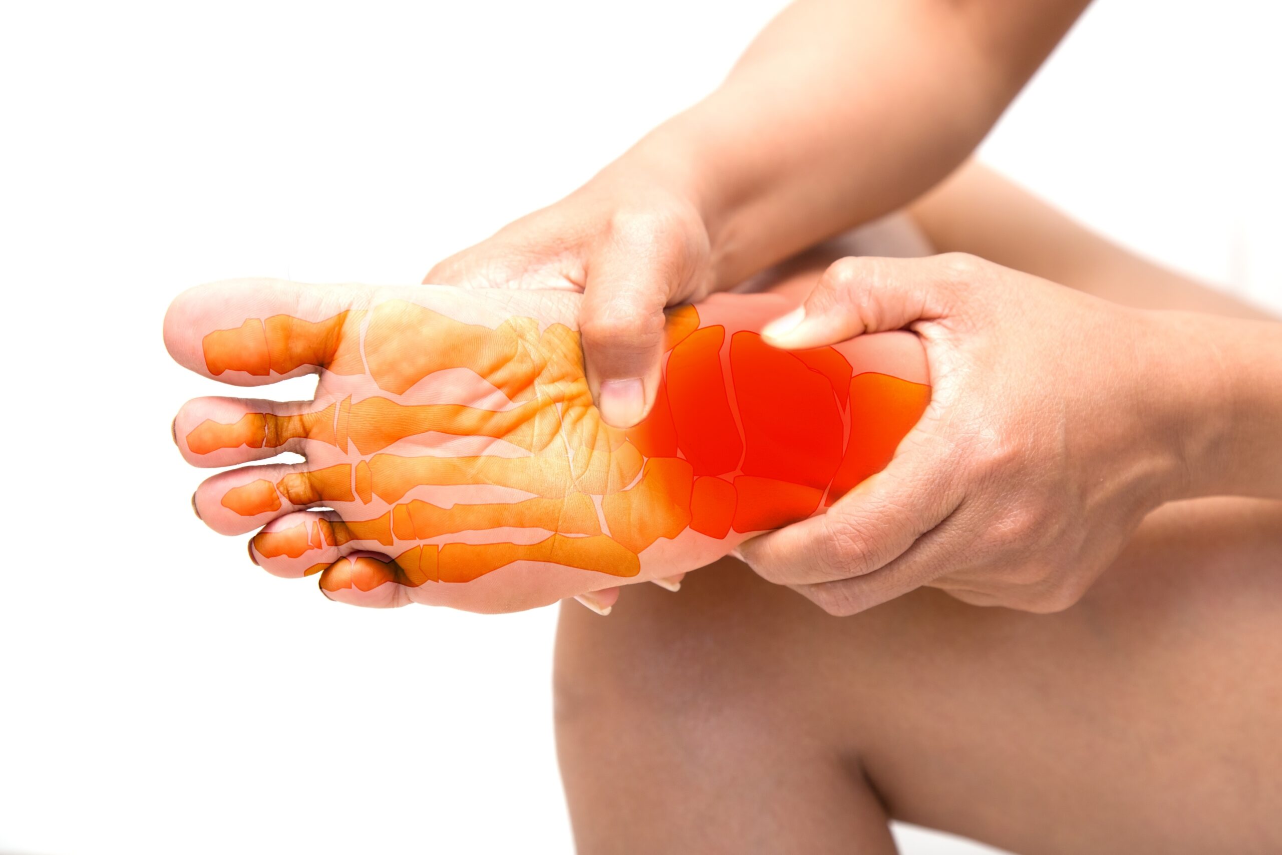
Most plantar fasciitis is diagnosed without a scan
A 2019 review in American Family Physician puts it simply: in most cases, clinical findings are enough to diagnose plantar fasciitis. Your doctor is listening for a typical story, which is pain under the heel that is worst with the first steps in the morning or after sitting, and then pressing for tenderness on the bottom of the foot just in front of the heel bone, where the fascia attaches.
The American Academy of Orthopaedic Surgeons (AAOS) says MRI and ultrasound are not routinely used to diagnose plantar fasciitis and are rarely ordered. If your story and exam are classic and you are early in treatment, skipping the scan is normal care, not a shortcut.
When imaging is worth doing
Imaging earns its place when it could change what happens next. The usual triggers are:
- Pain that has lasted more than about three months and is not responding. The same review says imaging may help find another diagnosis at that point.
- A story that does not fit. Burning or tingling, pain that builds with activity, pain at night or at rest, or tenderness in the wrong place.
- A sudden event. A pop or tearing feeling, or pain after a fall or a jump in training.
- Planning a procedure. A research review on heel pain imaging notes it is particularly useful in cases that do not respond to first-line care or when a more invasive treatment is being considered.
The American College of Radiology's guidance for chronic foot pain follows the same order. An X-ray is the usual first test. If the X-ray is normal or unclear and a soft tissue problem such as the fascia is suspected, ultrasound or MRI without contrast dye are both rated as usually appropriate next steps.
The three tests side by side
| Test | What it shows well | What it misses or overcalls | Typical role |
|---|---|---|---|
| X-ray | Bone: fractures, arthritis, heel spurs | Not sensitive for plantar fasciitis itself. Early stress fractures are often not visible. Spurs are common in people with no pain. | First test, mainly to rule out bone problems |
| Ultrasound | Fascia thickness and texture, fluid around the fascia, tears, increased blood flow on Doppler | Does not show the inside of bone | Confirming plantar fasciitis and measuring thickness. No radiation. |
| MRI | Fascia, bone marrow, nerves and surrounding soft tissue in one study | Can turn up findings that are also common in people without pain | Looking for a different or additional diagnosis |
X-ray: useful for bone, limited for fascia
The AAOS describes X-rays as useful for ruling out other causes of heel pain, such as fractures or arthritis. They will also show a heel spur if you have one, and this is where confusion starts.
People with long-lasting heel pain are more likely to have a spur. A pooled analysis found about 8.5 times the odds compared with people without heel pain. But the AAOS is clear that most people with heel spurs have no pain, and that spurs do not cause plantar fasciitis pain. The spur also stays on the X-ray after symptoms resolve. A spur on your report does not prove the diagnosis and is not a reason for surgery. We cover this in heel spur vs. plantar fasciitis.
One limit to know: a heel bone stress fracture often does not show on an early X-ray, so a normal film does not fully rule one out if your symptoms point that way.
Questions about your treatment options? Dr. Fox can help.
Book a ConsultationThis article is part of our Heel Pain Guide.
Ultrasound: measuring the fascia
Ultrasound looks directly at the fascia. The number most often quoted is thickness where the fascia meets the heel bone.
- In a 2009 meta-analysis of imaging studies, the fascia in people with chronic heel pain was on average 2.16 mm thicker than in people without it.
- A thickness greater than 4.0 mm is the usual cutoff taken to support the diagnosis. The same meta-analysis found people with heel pain were far more likely to be above it, although that estimate came from small studies and is imprecise.
- Other typical findings are a darker, less organized look to the fascia, fluid next to it, small calcium deposits and extra blood flow on Doppler.
How accurate is it? In a study of 77 patients that used MRI as the reference, ultrasound had 80% sensitivity and 88.5% specificity for plantar fasciitis. A systematic review of 10 studies concluded it was accurate and reliable compared with MRI. It also costs less and uses no radiation, which is why radiology reviews call it the first-line test for the fascia itself.
Two cautions about the thickness number. It does not predict your future: in a study that followed 174 patients for about 10 years, fascia thickness at the start had no effect on who recovered. And it is not a score you need to chase. A measurement is one piece of information next to your symptoms and exam.
MRI: the wider view
MRI shows the same thickening, along with signal changes inside the fascia and swelling in the nearby soft tissue and bone marrow. Its real value is in what else it can find. Doctors use it to look for a partial or complete fascia tear, a heel bone stress fracture, a pinched nerve such as Baxter's nerve or the tibial nerve in the tarsal tunnel, and problems at the Achilles attachment. Our guide to conditions mistaken for plantar fasciitis explains these look-alikes.
MRI can also show things that do not matter. For example, a muscle change sometimes read as a sign of Baxter's nerve trouble appears in 4% to 11% of the general population, with and without foot pain. An MRI report should be read against your symptoms, not on its own. Contrast dye is not usually needed for this question.
Does imaging change treatment?
Sometimes a great deal, sometimes not at all.
- It changes the plan when it finds something else. A stress fracture needs rest from impact, a tear changes what injections or procedures make sense, and a nerve problem calls for a different approach entirely.
- It supports the plan when it confirms a thick, abnormal fascia in someone considering a procedure.
- It changes little in a classic early case. Stretching, supportive shoes and time are the treatment whether the fascia measures 4.5 or 6 mm, and spur size does not guide care.
If your pain has dragged on, the step-by-step plan in plantar fasciitis that won't go away shows where a recheck with imaging fits. The heel pain guide links to every related topic.
What imaging is used before embolization?
Plantar fasciitis embolization (PFE) is a catheter procedure that reduces blood flow in the small abnormal vessels around a chronically painful fascia. Because it is a procedure, confirming the diagnosis first matters. There is no single published standard for which scan is required, and the studies have used different ones:
- A 2026 retrospective series of 12 patients required tenderness over the fascia plus a fascia thicker than 4 mm on MRI.
- A 2026 prospective study of 32 patients measured the fascia with ultrasound, and reported that average thickness fell from 5.2 mm to 3.7 mm six months after treatment.
- A 2026 series of 41 treated legs reviewed X-rays and found that having a heel spur made no difference to results.
During the procedure itself, the doctor takes angiogram pictures, which are X-ray images of the arteries with contrast dye, to find the abnormal vessels. That is part of the treatment, not a separate test to schedule.
Keep the evidence in perspective. PFE research is early. In a 2026 prospective series, 17 of 24 patients (70.8%) responded at six months, and in the 12-patient series, 8 of 11 treated (72.7%) did. These are small studies with no comparison group and no randomized trial. A thick fascia on a scan does not by itself mean you need a procedure.
What Dr. Fox orders depends on your history and exam. If you already have X-ray, ultrasound or MRI results, bring the reports and the images to your visit so tests are not repeated without reason. You can read more on our plantar fasciitis embolization page, in the plain-language explainer on TAME, TAE and PFE, and in the embolization guide.
When heel pain needs prompt attention
Do not wait for a routine appointment if you felt a pop or tearing in your heel or arch, cannot put weight on the foot, have heel pain after a fall, have deep pain at night or at rest that does not ease, have fever with a hot, red or swollen heel, or have new numbness. These situations often do need imaging, and soon.
Frequently Asked Questions
Can an X-ray show plantar fasciitis?
Not directly. An X-ray shows bone, so it is used to rule out problems such as a fracture or arthritis. It may show a heel spur, but most people with heel spurs have no pain and the spur is not the cause of plantar fasciitis pain.
What plantar fascia thickness is abnormal on ultrasound?
A thickness greater than 4.0 mm near the heel bone is the usual cutoff that supports plantar fasciitis. In pooled studies the fascia was on average 2.16 mm thicker in people with chronic heel pain than in people without it.
Is MRI better than ultrasound for plantar fasciitis?
For the fascia itself, ultrasound performs well and costs less. In one study it had 80% sensitivity and 88.5% specificity compared with MRI. MRI is more useful when the question is whether something else, such as a stress fracture, tear or nerve problem, is causing the pain.
Does a thicker fascia mean a worse outcome?
Not according to the longest follow-up study. Among 174 patients followed for about 10 years, fascia thickness and heel spurs at the start had no effect on who recovered.
Do I need an MRI before plantar fasciitis embolization?
There is no single published standard. Studies have used MRI, ultrasound or X-ray before treatment, and Dr. Fox decides what is needed after reviewing your history, exam and any scans you already have.
Keep Reading
- Plantar Fasciitis Embolization (PFE) in Manhattan
- Plantar Fasciitis When You Stand All Day at Work
- Plantar Fasciitis Surgery: Recovery, Success Rates and Risks
- TAME, TAE, PFE and Microembolization: Are They the Same Procedure?
- Plantar Fasciitis That Won't Go Away: A Practical Next-Step Plan
- Shockwave Therapy for Plantar Fasciitis Did Not Work. What Are Your Options?
- Heel Pain Guide: causes, treatments and PFE
Sources
- American Academy of Orthopaedic Surgeons, OrthoInfo: Plantar fasciitis and bone spurs
- Trojian T, Tucker AK, American Family Physician, 2019: Plantar fasciitis
- American College of Radiology, ACR Appropriateness Criteria: Chronic foot pain, 2025 update
- McMillan AM et al., J Foot Ankle Res, 2009: Diagnostic imaging for chronic plantar heel pain, systematic review and meta-analysis of 23 studies
- Draghi F et al., Insights Imaging, 2017: Imaging of plantar fascia disorders on radiography, ultrasound and MRI, review
- Sabir N et al., J Ultrasound Med, 2005: Clinical utility of sonography in diagnosing plantar fasciitis, 77 patients
- Radwan A et al., Int J Sports Phys Ther, 2016: Ultrasonography in diagnosing plantar fasciitis, systematic review of 10 studies
- Hansen L et al., Orthop J Sports Med, 2018: Long-term prognosis of plantar fasciitis, 174 patients followed about 10 years
- Tu P, American Family Physician, 2018: Heel pain, diagnosis and management
- Latt LD et al., Foot Ankle Orthop, 2020: Evaluation and treatment of chronic plantar fasciitis, expert review
- Chen JSC et al., J Foot Ankle Res, 2025: Fatty infiltration of abductor digiti minimi on MRI, systematic review of 4 studies
- Kumar N et al., J Vasc Interv Radiol, 2026: Superselective embolization for refractory plantar fasciitis, retrospective series of 12 patients
- Tonkaz M, Bekci T, Eur J Radiol, 2026: Embolization for plantar fasciitis, prospective single-center study of 32 patients
- Astani SA et al., J Vasc Interv Radiol, 2026: Plantar fasciitis embolization with resorbable embolics, 41 legs in 34 patients
- Gill S et al., J Vasc Interv Radiol, 2026: Transarterial embolization for plantar heel pain, 6-month prospective case series of 24 patients
Medically reviewed by David Fox, MD, FACS, RPVI. This article is general information, not a substitute for a medical evaluation.
Living with joint or tendon pain that hasn’t improved? Schedule a consultation with Dr. David Fox at Fox Vein & Vascular in Manhattan to find out whether embolization is right for you. Call (212) 362-3470 or request an appointment online.