Plantar fasciitis is the most common reason for pain under the heel, so it is often the first label a sore heel gets. Most of the time the label is right. But several other conditions hurt in nearly the same spot, and they do not respond to plantar fasciitis treatment. If your heel has not improved after months of doing everything right, it is fair to ask whether the diagnosis is correct. This guide covers the main look-alikes, how their pain differs and which tests sort them out.
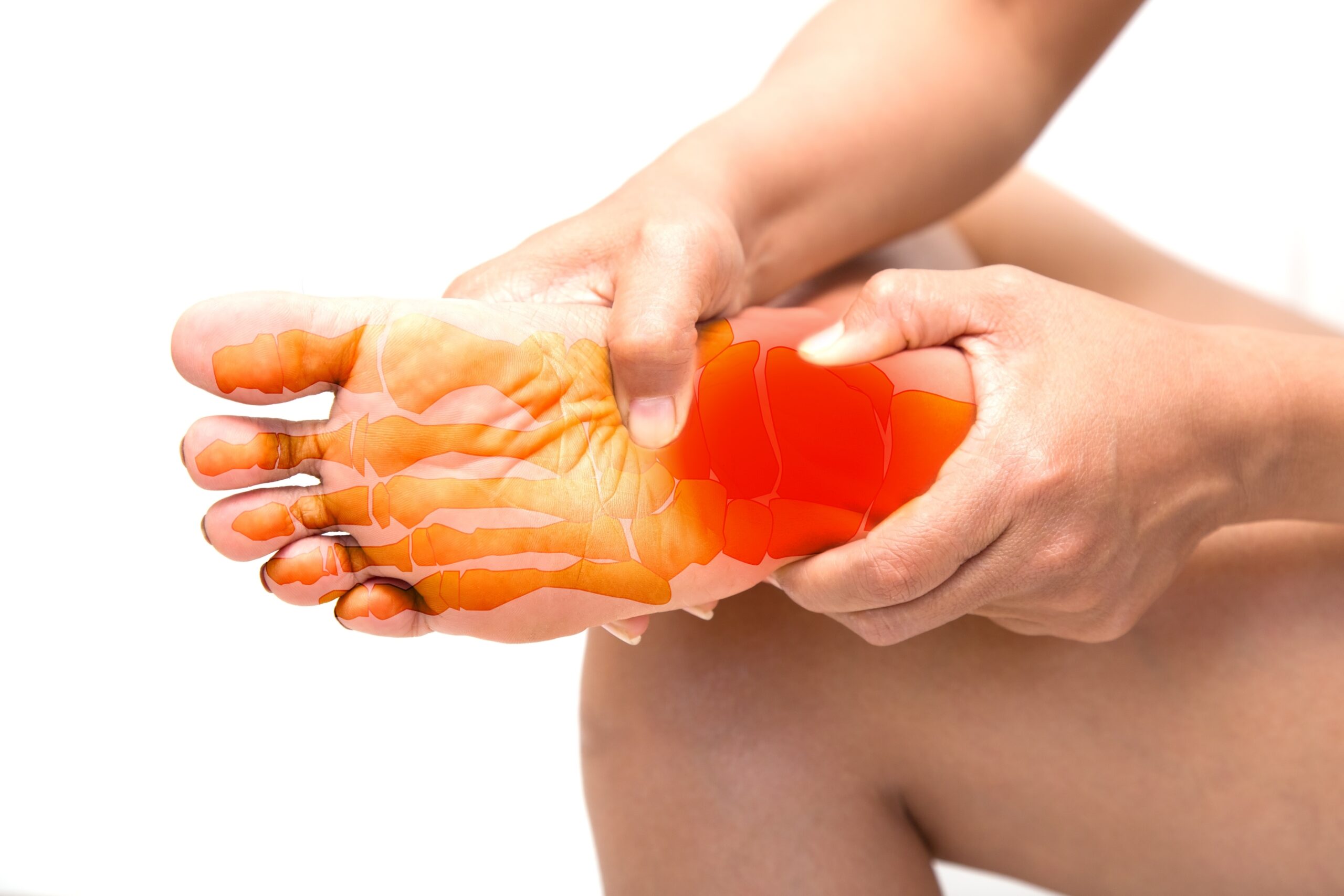
What typical plantar fasciitis feels like
It helps to start with the usual pattern. Plantar fasciitis causes pain on the inner underside of the heel that is worst with the first steps in the morning or after sitting, eases as you move, and often returns after long periods on your feet. Pressing on the inner edge of the heel bone is tender. There is usually no numbness, tingling or burning, and no swelling or redness. Our page on morning heel pain explains the pattern in more detail.
When your symptoms do not match that description, one of the conditions below may be involved. How often does that happen? In one review of 250 patients seen for pain under the heel at a single foot clinic, 53.2% had plantar fasciitis alone, 14.8% had heel fat pad atrophy, 9.2% had both, 1.6% had a plantar fascia rupture and 0.8% had a nerve problem. Other clinics report different mixes, but the point holds: a meaningful share of heel pain is something else.
The look-alikes at a glance
| Condition | How the pain differs | What helps confirm it |
|---|---|---|
| Baxter's nerve entrapment | Burning pain on the inner underside of the heel, sometimes with tingling | Exam, nerve tests (EMG), high-resolution ultrasound, MRI |
| Tarsal tunnel syndrome | Numbness, burning or tingling in the heel, inner ankle and sole | Tapping over the nerve at the inner ankle (Tinel sign), MRI |
| S1 or other low back nerve root | Heel pain or numbness that may come with leg or back symptoms, though back pain can be absent | Neurologic exam, spine imaging if needed |
| Heel fat pad syndrome | Deep, bruise-like pain in the center of the heel, worse barefoot or on hard floors | Firm pressure on the center of the heel, ultrasound |
| Calcaneal stress fracture | Pain that builds after an increase in activity, hurts during weight bearing and later at rest | Squeezing the heel from the sides, MRI, CT or bone scan |
| Insertional Achilles tendinopathy | Pain at the back of the heel, often with shoe pressure | Exam, X-ray for spurs at the back of the heel |
| Plantar fascia tear | Sudden tearing or pop, often after a steroid injection | Exam, ultrasound or MRI |
| Inflammatory arthritis | Heel pain in both feet, or with other joint, back or skin symptoms | Blood tests, ultrasound, rheumatology evaluation |
Nerve problems: Baxter's nerve, tarsal tunnel and the low back
Nerve pain has its own vocabulary. People describe burning, tingling, electric or numb sensations, which are not typical of plantar fasciitis.
Baxter's nerve entrapment. Baxter's nerve, also called the inferior calcaneal nerve, is the first branch of the lateral plantar nerve. It can be squeezed as it passes under the inner heel, very close to where plantar fasciitis hurts. How common this is remains uncertain. One 2025 review estimated it accounts for up to 20% of chronic heel pain, while the 250-patient clinic series above found nerve causes in under 1%. MRI can show changes in a small foot muscle the nerve supplies, but a 2025 systematic review found those changes in 4% to 11% of the general population, so the scan alone does not settle it.
Tarsal tunnel syndrome. Here the tibial nerve is compressed behind the inner ankle bone. It causes numbness, burning and tingling in the heel, inner ankle and sole. Doctors look for symptoms that are reproduced by tapping over the nerve. MRI is the preferred imaging test because it can show what is pressing on the nerve.
A pinched nerve in the low back. The nerve roots that supply the heel, including S1, start in the lower spine. American Family Physician advises doctors to consider a lower back nerve root problem in heel pain even when there is no back pain. Clues include symptoms that run down the back of the leg, change with sitting or bending, or come with weakness.
Bone and cushion problems: stress fracture and fat pad syndrome
Calcaneal stress fracture. This is a small crack in the heel bone from repeated load. The heel bone is the second most common site for a stress fracture in the foot. Pain usually begins after an increase in weight-bearing activity or a switch to a harder surface. At first it hurts only with activity, and later at rest as well. Pain when the heel is squeezed from both sides suggests the diagnosis. Plain X-rays often miss it early, so an MRI, CT or bone scan may be needed. This is one you do not want to walk through.
Heel fat pad syndrome. The fat pad is the natural cushion under your heel bone. When it thins or is injured, you get a deep, bruise-like ache in the middle of the heel, not at the inner edge. It is worse barefoot, on hard floors and after long standing. In the clinic series above, fat pad atrophy more often affected both heels and caused pain at night. A 2022 review suggested it may be the second most common cause of pain under the heel, and also found very little research on how best to treat it. Thinning on ultrasound can support the diagnosis. Prior steroid injections, age and higher body weight are thought to contribute.
Questions about your treatment options? Dr. Fox can help.
Book a ConsultationThis article is part of our Heel Pain Guide.
Tendon and fascia injuries
Insertional Achilles tendinopathy. The key difference is location. This pain is at the back of the heel, where the Achilles tendon attaches, not underneath. The AAOS notes that bone spurs often form at the back of the heel with this condition and that it tends to hurt when you wear shoes. An MRI is not needed to diagnose it.
Plantar fascia tear. A tear is different from the gradual wear of plantar fasciitis. In a series of 765 patients treated for plantar fasciitis, 51 were diagnosed with a rupture, and 44 of those ruptures were associated with a corticosteroid injection. About two thirds of those 44 patients described a sudden tearing at the heel. A separate study of 286 patients found that 33 of the 35 with a rupture had received steroid injections. If your heel pain changed suddenly after an injection, with a pop, new arch pain or bruising, tell your doctor. Our article on cortisone shots that did not work covers what comes next.
Inflammatory arthritis
Conditions such as psoriatic arthritis, ankylosing spondylitis, rheumatoid arthritis and gout can inflame the heel where tendons and fascia attach. American Family Physician advises that when heel pain affects both feet, an underlying body-wide condition should be ruled out. Other clues are morning stiffness in the back or other joints, a swollen toe or finger, psoriasis, or eye or bowel inflammation.
The diagnosis is not always simple. In a small ultrasound study of 27 people with spondyloarthritis and tender heels, fewer than half had signs of true inflammation at the attachment point. A rheumatologist usually makes this call, using blood tests and imaging together.
Which tests sort these out
- History and exam. Where exactly it hurts, when, and what it feels like does most of the work.
- X-ray. Useful for ruling out fractures and arthritis and for seeing spurs. See heel spur vs. plantar fasciitis for what a spur does and does not mean.
- Ultrasound. Shows fascia thickness, tears, fat pad thinning and inflammation.
- MRI. Not routine for plantar fasciitis, but ordered when a stress fracture, tear, nerve compression or another problem is suspected.
- Nerve studies and blood tests. Used when nerve entrapment or inflammatory arthritis is on the list.
Not sure who should run this workup? See which doctor to see for plantar fasciitis.
Red flags that need prompt care
See a doctor soon, or go to urgent care, if you have:
- A sudden pop or tearing with new swelling or bruising
- Inability to put weight on the foot
- A hot, red, swollen heel, or fever
- Numbness or weakness that is spreading, or new trouble controlling your bladder or bowels along with back or leg symptoms
- Pain that wakes you at night or is constant at rest, especially with unexplained weight loss
- A sore or wound on the heel if you have diabetes
Why this matters before embolization
Plantar fasciitis embolization (PFE) reduces abnormal blood flow around an inflamed plantar fascia. It has only been studied for plantar fasciitis. It will not heal a stress fracture, free a trapped nerve, rebuild a fat pad or treat arthritis. That is why Dr. Fox, a board-certified vascular surgeon, considers PFE only once the diagnosis is confirmed, which may mean imaging or a visit with a foot specialist first.
Even with the right diagnosis, PFE research is early. The studies so far are small series without comparison groups, such as a 2026 study in which 17 of 24 people (70.8%) responded at 6 months, and there is no randomized trial. You can read more on our plantar fasciitis embolization page, in the heel pain guide and in the wider embolization guide.
Frequently Asked Questions
How do I know if my heel pain is a nerve problem and not plantar fasciitis?
Burning, tingling, electric or numb sensations point toward a nerve. Plantar fasciitis usually causes a sharp or aching pain with first steps and does not cause numbness.
Can a stress fracture feel like plantar fasciitis?
Yes, early on. A stress fracture tends to hurt more during weight-bearing activity, can progress to pain at rest, and is tender when the heel is squeezed from the sides. Early X-rays can be normal, so an MRI or other scan may be needed.
Do I need an MRI for heel pain?
Usually not. The AAOS says MRI is not routinely used for plantar fasciitis, but a doctor may order one if another problem is suspected or the pain is not improving as expected.
Should I have embolization if my diagnosis is uncertain?
No. PFE has only been studied for plantar fasciitis, so the diagnosis should be confirmed first. Dr. Fox may ask for imaging or an evaluation by a foot specialist before considering it.
Keep Reading
- Plantar Fasciitis Embolization (PFE) in Manhattan
- Your Cortisone Shot for Plantar Fasciitis Did Not Work. What Now?
- Heel Pain at Night, Burning Heel Pain and Pain After Sitting: What Each Pattern Suggests
- Heel Spur vs. Plantar Fasciitis: What Is the Difference?
- Plantar Fasciitis Embolization for Runners and Athletes: What to Know
- Plantar Fasciitis Embolization vs. Radiofrequency Ablation and Topaz: How They Compare
- Heel Pain Guide: causes, treatments and PFE
Sources
- Tu P, Am Fam Physician, 2018: Heel pain, diagnosis and management
- Lareau CR et al., J Am Acad Orthop Surg, 2014: Plantar and medial heel pain, diagnosis and management
- Yi TI et al., Ann Rehabil Med, 2011: Clinical characteristics of the causes of plantar heel pain, review of 250 patients
- Chang AH et al., J Foot Ankle Res, 2022: Heel fat pad syndrome, scoping review
- Tedeschi R, Neurol Sci, 2025: Baxter's nerve, the hidden culprit of chronic heel pain (narrative review)
- Chen JSC et al., J Foot Ankle Res, 2025: Baxter's neuropathy and fatty infiltration of the abductor digiti minimi on MRI, systematic review
- Fortier LM et al., Orthop Rev, 2022: An update on posterior tarsal tunnel syndrome
- American Academy of Orthopaedic Surgeons, OrthoInfo: Stress fractures of the foot and ankle
- American Academy of Orthopaedic Surgeons, OrthoInfo: Achilles tendinitis
- American Academy of Orthopaedic Surgeons, OrthoInfo: Plantar fasciitis and bone spurs
- Acevedo JI, Beskin JL, Foot Ankle Int, 1998: Plantar fascia rupture associated with corticosteroid injection, 765 patients
- Lee HS et al., Foot Ankle Int, 2014: Risk factors affecting chronic rupture of the plantar fascia, 286 patients
- Felbo SK et al., J Clin Med, 2022: Ultrasound of tender heel entheses in peripheral spondyloarthritis, 27 patients
- Gill S et al., J Vasc Interv Radiol, 2026: Transarterial embolization for plantar heel pain, 6-month prospective case series of 24 patients
Medically reviewed by David Fox, MD, FACS, RPVI. This article is general information, not a substitute for a medical evaluation.
Living with joint or tendon pain that hasn’t improved? Schedule a consultation with Dr. David Fox at Fox Vein & Vascular in Manhattan to find out whether embolization is right for you. Call (212) 362-3470 or request an appointment online.