Many patients walk into a specialist’s office carrying an X-ray report and a deep sense of frustration. They have been told they “have arthritis,” but that label rarely explains why their knee hurts on some days and not others. A basic diagnosis often fails to answer the most important questions: why is the pain happening now, and what should be done next?
Getting a clear diagnosis of knee osteoarthritis requires more than just a quick glance at an imaging scan. True diagnostic clarity comes from understanding the relationship between the physical structure of your knee, the symptoms you experience, and the underlying inflammation driving the discomfort.
When you understand how a diagnosis is properly made, you gain the power to make informed decisions about your care. This process moves beyond a simple anatomical label, focusing instead on identifying the precise factors causing your pain so you can find meaningful relief without unnecessary procedures.
Why Knee Arthritis Diagnosis Isn’t Always Straightforward
Diagnosing knee pain is rarely as simple as matching a picture to a problem. The human body is complex, and the way it registers pain does not always align with what appears on a screen.
Why imaging alone doesn’t explain your pain
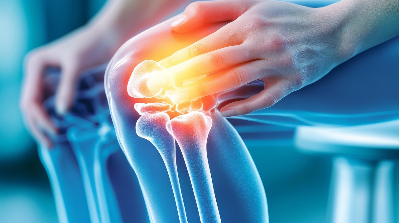
A common misconception is that an X-ray serves as a definitive answer for knee pain. While imaging provides a useful map of your bone structure, it cannot show pain. It cannot show the exact source of your daily discomfort or the complex vascular changes happening inside the joint. Using imaging as the sole diagnostic tool often leads to incomplete conclusions about why knee osteoarthritis causes pain.
Why two people with the same X-ray can feel completely different
Two patients can have identical X-rays showing bone-on-bone arthritis. One patient might struggle to walk to the mailbox, while the other plays 18 holes of golf with minimal discomfort. The severity of structural joint damage does not reliably predict the severity of a patient’s symptoms. This discrepancy highlights why treating the X-ray instead of the patient often leads to poor clinical outcomes.
The difference between structural damage and active pain
Structural damage refers to the physical loss of cartilage over time. Active pain is typically driven by biological processes, including the vascular theory of osteoarthritis. As cartilage wears down, the body often responds by forming abnormal, leaky blood vessels in the joint lining, leading to swelling, nerve irritation, and active pain. Differentiating between old structural changes and new, active inflammation is critical for an accurate diagnosis.
The First Step: Understanding Your Symptoms
Your daily experience with pain provides the most valuable diagnostic clues. A thorough evaluation always begins with a detailed conversation about how your knee actually feels.
How pain patterns help guide diagnosis
Pain rarely behaves randomly. A specialist will want to know exactly when your knee hurts, how long the pain lasts, and what makes it worse. Sharp pain when walking down stairs suggests a different problem than a constant, dull ache that keeps you awake at night. These distinct patterns help pinpoint the primary drivers of your knee arthritis symptoms.
What stiffness, swelling, and timing can reveal
Morning stiffness that improves after a few minutes of movement is a hallmark sign of osteoarthritis. In contrast, a knee that swells dramatically after a specific activity points toward active inflammation or a potential mechanical issue. The timing of these symptoms gives doctors insight into whether the primary issue is mechanical friction, fluid buildup, or chronic joint pain triggered by an inflammatory response.
Why your daily limitations matter more than you think
Doctors need to know how your knee affects your life. If you have stopped walking your dog, taking the subway, or exercising because of your knee, those limitations provide a baseline for measuring treatment success. The goal of chronic joint pain treatment is not to make an X-ray look better; it is to restore your ability to live your life comfortably.
Physical Examination: What Doctors Are Actually Looking For
A physical exam tells a specialist things an image never could. It allows the doctor to feel how the joint moves and identify exactly where the tissues are irritated.
Range of motion and joint restriction
During the exam, the doctor will move your knee through its full range of motion. They are feeling for a grinding sensation known as crepitus, testing the stability of the ligaments, and noting any points where the joint catches or physically stops moving. Restricted motion often indicates tight joint capsules or bone spurs physically blocking the path.
Swelling, tenderness, and joint alignment
A specialist will gently press around the knee to identify specific areas of tenderness. They will also look at the overall alignment of your leg. If your knee bows inward or outward, it places abnormal stress on specific compartments of the joint. Evaluating the presence of spongy, fluid-filled swelling helps determine the extent of synovial inflammation.
Identifying mechanical vs inflammatory pain
Mechanical pain typically happens during specific movements—like standing up from a chair—and stops when the movement stops. Inflammatory pain behaves differently. It often throbs even when you are resting, causes the knee to feel warm to the touch, and creates a generalized feeling of pressure. Understanding how inflammation leads to chronic joint pain helps direct the right therapeutic approach.
Imaging Tests Used to Diagnose Knee Arthritis
While imaging should not be the only tool used, it remains a vital part of the diagnostic puzzle when utilized correctly.
X-rays and what they can (and can’t) show
X-rays are excellent at showing bone. They can reveal narrowing joint spaces, bone spurs, and structural deformities. However, they cannot show cartilage, ligaments, tendons, or the inflamed joint lining. An X-ray is a helpful starting point, but it provides an incomplete picture of a living, moving joint.
MRI and when it’s actually useful
Magnetic Resonance Imaging (MRI) provides a detailed view of the soft tissues in the knee. It is particularly useful if a doctor suspects a meniscus tear, ligament damage, or severe cartilage loss. However, MRIs are highly sensitive and often highlight minor abnormalities that are entirely unrelated to the patient’s actual pain, which can sometimes lead to unnecessary anxiety.
Ultrasound and evaluating inflammation or fluid
Ultrasound is an incredibly practical tool because it allows the doctor to look inside the joint while it is moving. It is highly effective at identifying fluid buildup, swollen tissues, and abnormal blood flow. In a diagnostic vascular laboratory setting, understanding how ultrasound detects chronic conditions allows specialists to accurately target the active sources of inflammation.
Why Imaging Doesn’t Always Match Your Symptoms
The disconnect between imaging results and physical symptoms is one of the most confusing aspects of knee arthritis for patients to navigate.
Mild arthritis with severe pain
Some patients have X-rays that look relatively normal, showing only mild signs of wear. Yet, they experience debilitating pain. This often happens because the joint lining is severely inflamed, packed with abnormal nerve fibers and blood vessels that are hyper-sensitive to any movement.
Advanced arthritis with minimal symptoms
Conversely, older adults often have X-rays that look terrible—showing advanced, bone-on-bone disease—yet they walk without a limp. Their bodies have adapted to the structural changes over decades, and they lack the active inflammatory response that triggers severe pain signals.
The role of inflammation and blood flow
The missing link between imaging and symptoms is often vascular. When cartilage breaks down, the joint attempts to heal itself by growing new blood vessels. These vessels are abnormal and bring excess inflammatory cells and nerve endings into the joint. Understanding how blood flow contributes to knee pain and synovial inflammation is critical to understanding why your knee actually hurts.
Identifying Inflammation as a Pain Driver
Recognizing inflammation as the primary culprit behind your knee pain changes the entire diagnostic landscape.
Signs inflammation is contributing to your symptoms
If your knee frequently feels tight, warm, or swollen, inflammation is at work. If you experience a persistent throbbing ache at the end of the day or pain that wakes you up at night, these are classic indicators that the joint lining is actively inflamed and irritated.
How vascular changes affect joint pain
Chronic inflammation physically alters the environment inside your knee. Based on the vascular theory of osteoarthritis, the abnormal blood vessels that form in an arthritic knee maintain a constant state of swelling. These vessels supply the sensory nerves with inflammatory chemicals, keeping the pain dial turned all the way up.
Why this changes how treatment is approached
If your pain is driven by inflammation and abnormal blood flow rather than pure structural breakdown, you do not necessarily need to replace the joint. This realization shifts the focus toward inflammation-based pain treatment, allowing doctors to target the exact biological mechanism causing the discomfort.
Questions about your treatment options? Dr. Fox can help.
Book a ConsultationRuling Out Other Causes of Knee Pain
Knee pain is not always arthritis. A precise diagnosis requires ruling out other conditions that mimic osteoarthritis symptoms.
Injury-related knee pain vs arthritis
A sudden twist or fall can tear a meniscus or sprain a ligament, leading to acute pain. While an older injury can eventually lead to arthritis, acute sports injuries require a different diagnostic approach and a specific type of sports injury treatment.
Tendonitis and overuse conditions
Repetitive stress can irritate the tendons surrounding the knee, causing conditions like patellar tendonitis. This type of pain is usually localized to the front of the knee and worsens with activities like jumping or running. It is an issue of the soft tissue, not the joint itself.
Circulation-related leg pain and PAD
Sometimes, pain felt in the lower extremities is not coming from the joints at all. Poor circulation, particularly peripheral arterial disease (PAD), can cause cramping and pain in the legs during walking. A thorough specialist will evaluate the vascular health of the leg to distinguish leg pain vs vein pain from joint-related issues.
When Knee Arthritis Is Confirmed
Receiving a confirmed diagnosis of knee osteoarthritis is a starting point, not a sentence.
Understanding severity vs symptoms
A doctor grading your arthritis as “moderate” or “severe” is describing the structural appearance on an X-ray. It is vital to remember that you are treating the symptoms, not the grade. Your treatment plan should be based on how much the arthritis limits your life, not just how it looks on a screen.
Why diagnosis doesn’t automatically mean surgery
Many patients assume that an arthritis diagnosis means a joint replacement is inevitable. This is false. A vast majority of patients successfully manage their symptoms and maintain their mobility using targeted, non-surgical approaches. Finding alternatives to knee replacement is entirely possible when you understand the root cause of the pain.
What most patients are told—and what’s often missing
Patients are frequently told to “lose weight, take ibuprofen, and come back when it’s time for surgery.” What is missing from this conversation is a nuanced explanation of how to avoid knee replacement by targeting the abnormal blood vessels and inflammation driving the daily pain.
How Diagnosis Guides Treatment Decisions
A precise diagnosis allows a specialist to build a logical, customized treatment plan.
Matching treatment to symptom patterns
If a patient’s primary complaint is mechanical stiffness, physical therapy to strengthen the surrounding muscles might be the best first step. If the primary symptom is severe, fluid-filled swelling, the focus must shift to reducing the synovial inflammation causing the fluid buildup.
When conservative treatments still make sense
For many people, weight management, targeted physical therapy, and bracing offer substantial relief. When combined with a clear understanding of what activities to modify, these conservative measures remain the cornerstone of how to treat knee osteoarthritis without surgery.
When to consider minimally invasive options
When conservative measures fail to provide relief, the next logical step is not always a major operation. By utilizing the best non-surgical treatment for knee osteoarthritis, such as targeting the inflammatory blood supply to the joint lining, patients can find significant relief with minimal downtime.
When to Consider Advanced Evaluation or Treatment
There comes a point when basic interventions are no longer enough, and a more advanced evaluation is necessary.
Persistent pain despite therapy or injections
If you have completed months of physical therapy and tried cortisone or gel injections with only temporary or no relief, your current treatment plan is no longer matching your condition. This is a clear signal that the underlying inflammation needs to be addressed differently.
Symptoms that are limiting daily activity
When knee pain dictates your schedule—preventing you from sleeping, working, or enjoying your family—it is time to seek an advanced evaluation. You should not have to accept severe limitations as a normal part of aging.
Patients not ready for knee replacement
Many patients are told they need a knee replacement but are either too young, medically unfit, or simply unwilling to undergo a major surgery and lengthy rehab. For these individuals, exploring advanced minimally invasive options like genicular artery embolization (GAE) provides a powerful alternative. Discussing whether GAE is right for you can open doors to relief that bridges the gap between basic injections and total joint replacement.
What to Expect From a Knee Arthritis Evaluation
A proper evaluation at a specialty clinic looks different from a standard five-minute doctor’s visit.
Consultation and symptom review
The process begins with a conversation. The doctor will listen to your history, review any previous treatments you have tried, and ask detailed questions about your pain patterns. You will have the time to explain exactly how the knee feels.
Imaging and diagnostic planning
If new imaging is required, the doctor will explain exactly what they are looking for. They may use ultrasound in the office to look for active inflammation or order a specific type of MRI if a structural issue is suspected.
Personalized treatment discussion
Once the clinical exam and imaging are combined, the specialist will sit down with you to explain the findings. They will clearly outline the relationship between your structural arthritis and your active inflammation, discussing the most effective knee arthritis pain treatment options available for your specific case.
Next Step: Understanding What Your Diagnosis Means
Diagnosis should lead to clarity, not fear. A label of knee arthritis simply means your joint is changing, but it does not mean you have to surrender your quality of life. By understanding the difference between structural wear and active inflammation, you can take control of your health and pursue treatments that actually address the source of your pain.
If you are tired of generic answers and ready for a clear, actionable plan, it is time to explore effective non-surgical knee pain relief tailored to your unique condition.