If you have ever visited a doctor for leg pain or swelling, you might have expected an X-ray or maybe an MRI. But when you walk into a Manhattan vein clinic complaining of bulging veins or heaviness in your legs, the most important tool in the doctor’s arsenal isn’t a giant scanning machine—it is a handheld wand known as a Duplex Ultrasound.
For many patients, the term “ultrasound” brings to mind images of pregnancy scans. While the technology is similar, the application in vascular medicine is quite different. In the hands of a skilled vein specialist in Manhattan, Duplex Ultrasound is the gold standard for diagnosing vein disease. It is the map that guides every decision, from the first diagnosis to the final treatment.
If you are searching for a vein specialist near me or wondering why your doctor is recommending an ultrasound for your spider veins, this guide is for you. We will dive deep into how this non-invasive technology works, what it reveals about your health, and why it is the critical first step in effective vein care treatment in Manhattan. For more detailed treatment information, visit the Manhattan Vein Treatment page here.
What is Duplex Ultrasound?
To understand how a varicose vein doctor in Manhattan diagnoses your condition, you first need to understand the tool they are using. The term “Duplex” refers to the fact that this ultrasound combines two different types of sensing technologies into one screen.
1. Traditional (B-Mode) Ultrasound
This is the “picture” part of the exam. It uses sound waves that bounce off blood vessels and tissues to create a grayscale image of the anatomy. It allows the doctor to see the structure of your veins, their size, and whether there are any blockages like clots.
2. Doppler Ultrasound
This is the “flow” part of the exam. Doppler technology bounces sound waves off moving objects—in this case, your red blood cells. By measuring how the sound waves change pitch as they bounce back, the machine can calculate the speed and direction of blood flow. On the screen, this is often visualized as colors (red and blue) overlaying the grey image.
When combined, these two modes allow a chronic vein condition specialist near me to see the vein structure and the blood flow simultaneously. This provides a complete functional picture of your circulatory health.
Why Visual Inspection Isn’t Enough
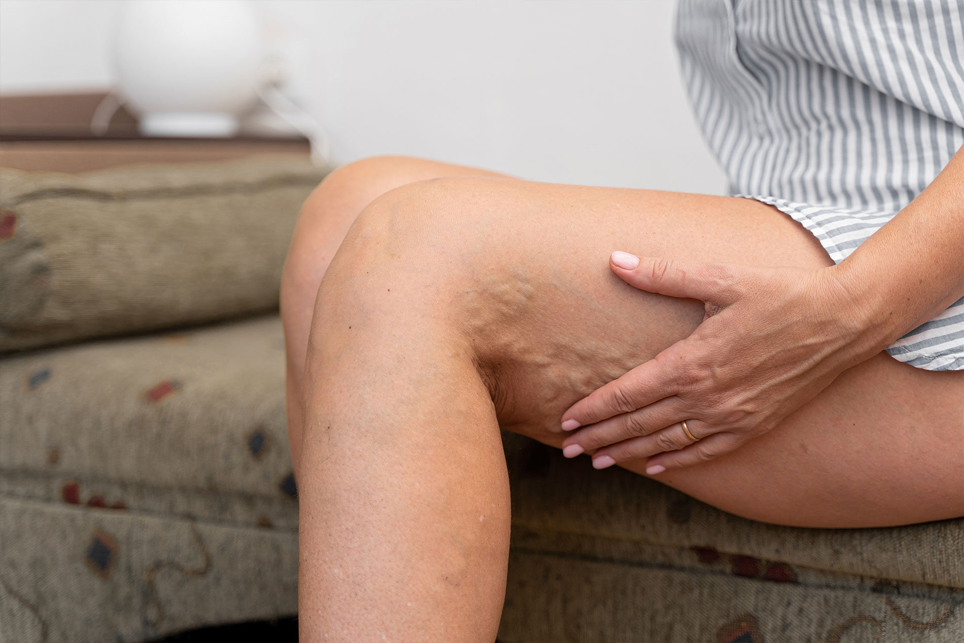
You might look down at your legs and think the problem is obvious. You see the twisted, purple ropes of varicose veins. You see the leg swelling and veins popping out after a long day. Why does the doctor need a fancy scan to tell you what you already know?
The reason is that what you see on the surface is often just the tip of the iceberg. The visible veins are usually secondary branches. The root cause of the problem is typically hiding deeper under the skin, in veins you cannot see with the naked eye, such as the Great Saphenous Vein or the Small Saphenous Vein.
Without an ultrasound, a doctor is just guessing. Treating surface veins without fixing the underlying “feeder” veins is a recipe for failure. The veins will simply return, often worse than before. A Best vein doctor near me uses ultrasound to find the source of the leak, ensuring that the treatment is permanent.
The Condition We Are Looking For: Venous Reflux
The primary goal of a venous ultrasound is to check for “Venous Reflux,” also known as Chronic Venous Insufficiency (CVI).
How Healthy Veins Work
In a healthy leg, veins have a tough job. They have to push blood upward, against gravity, back to the heart. To help with this, they have tiny, one-way valves inside them. When your muscles contract (like when you walk), blood is squeezed up. When the muscle relaxes, the valves snap shut to stop the blood from falling back down.
What Happens in Vein Disease
In patients with vein disease, these valves become weak or damaged. They don’t close all the way. Consequently, when gravity pulls down, the blood leaks backward through the open valve. This backward flow is called reflux.
Reflux causes blood to pool in the lower leg, increasing pressure in the veins. This pressure is what eventually causes the vein walls to stretch and bulge, creating varicose veins and leading to symptoms like vein pain and heaviness.
What to Expect During the Exam
If you have booked an appointment for vein treatment near me, knowing what to expect during the diagnostic scan can ease your anxiety. The process is painless, non-invasive, and radiation-free.
Step 1: Preparation
There is very little preparation needed. You should wear loose clothing or bring shorts, as the technician needs access to your entire leg, from the groin to the ankle. Hydration helps, as well-hydrated veins are easier to visualize.
Step 2: Positioning
Unlike many medical exams where you lie flat, a venous reflux study is often performed while you are standing up or sitting with your legs dangling.
- Why stand? Gravity is the key. If you lie flat, the pressure in your veins drops, and reflux might not happen. A vein specialist near me needs to see how your veins struggle against gravity to get an accurate diagnosis.
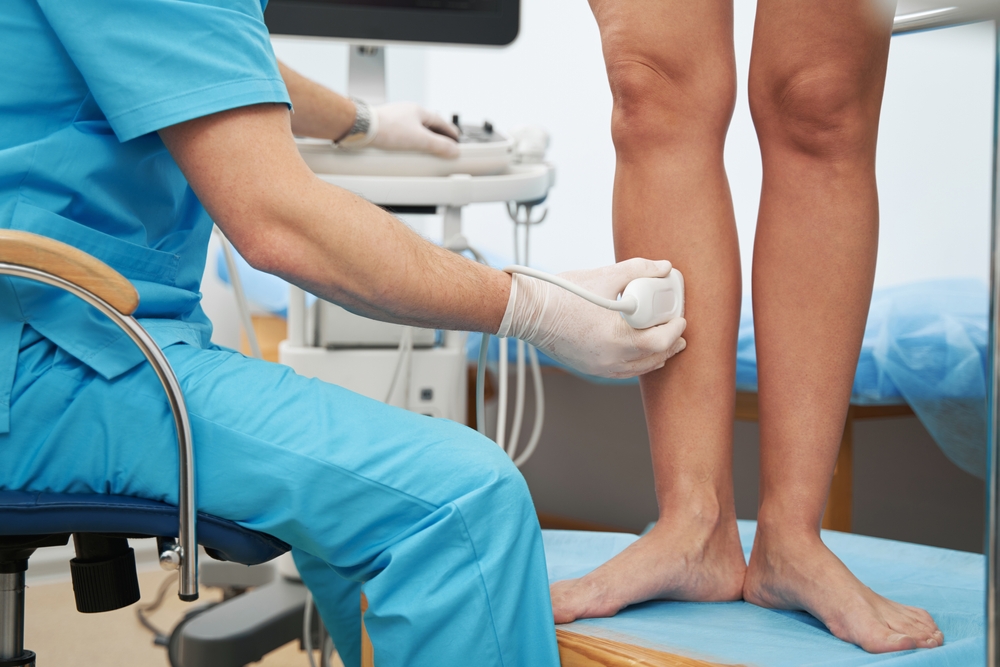
Step 3: The Scan
The technologist applies warm gel to your leg and moves the transducer wand over the skin. You will feel firm pressure but no pain. You might hear “whooshing” sounds from the machine—that is the sound of your blood flowing!
Step 4: The Squeeze (Augmentation)
This is the unique part of a vein check. The technician will squeeze your calf muscle firmly and then let go quickly.
- The Squeeze: Pushes blood up (mimicking walking).
- The Release: The technician watches the screen immediately after letting go. In a healthy vein, flow stops instantly as valves close. In a diseased vein, they will see (and hear) blood rushing backward.
Interpreting the Results: What the Colors Mean
During the exam, you will likely look at the screen. You will see flashes of blue and red. It is a common misconception that blue means “vein” and red means “artery.” In ultrasound physics:
- Red usually indicates blood flowing towards the probe.
- Blue usually indicates blood flowing away from the probe.
A vein specialist in Manhattan analyzes these colors to determine direction. If the probe is at the top of your calf and the blood should be moving up, but the color indicates it is moving down, that is visual proof of reflux.
The doctor also measures the time the reflux lasts.
- < 0.5 seconds: Generally considered normal valve closure time.
- > 0.5 seconds: Indicates significant reflux (valve failure).
This specific timing is crucial for insurance purposes. Most insurance providers require proof of reflux lasting longer than 0.5 seconds to cover procedures for vein symptoms in legs.
Ruling Out Deep Vein Thrombosis (DVT)
While the primary focus of a vein clinic visit is often chronic issues like spider veins or aching legs, the ultrasound serves a critical safety function: ruling out Deep Vein Thrombosis (DVT).
DVT is a serious condition where a blood clot forms in the deep veins of the leg. If a piece of this clot breaks off, it can travel to the lungs and cause a pulmonary embolism, which can be fatal.
Symptoms of DVT include:
- Sudden, intense leg swelling and veins feeling tender.
- Redness and warmth in one leg.
- Pain that feels like a cramp but doesn’t go away.
Because the symptoms of DVT can mimic severe varicose veins or muscle injuries, a Manhattan vein clinic will always check the deep system first. On the ultrasound, a healthy vein is compressible—if the technician presses on it with the wand, the vein walls touch. If there is a clot inside, the vein will not collapse. This “compressibility test” is the definitive way to diagnose a clot.
Learn more about DVT and vein conditions here.
Mapping the Anatomy for Treatment
Once the doctor has confirmed reflux and ruled out clots, the ultrasound becomes a mapping tool. No two people have the exact same vein anatomy. Your veins are as unique as your fingerprints.
A chronic vein condition specialist near me creates a “vein map.” This is a diagram of your leg that marks:
- Reflux Points: Exactly where the valves have failed.
- Perforator Veins: Connections between the deep and superficial systems that might be leaking.
- Tortuosity: Areas where the vein is too twisted to pass a catheter through.
- Nerve Proximity: Identifying where sensitive nerves run close to the vein to avoid injury during treatment.
This map is essential for planning procedures like Endovenous Laser Therapy (EVLT). It tells the doctor exactly where to insert the fiber, where to start the laser energy, and where to stop.
Diagnosis of Pelvic Congestion Syndrome
Sometimes, vein pain in the legs originates much higher up. Pelvic Congestion Syndrome (PCS) involves varicose veins in the pelvis and lower abdomen. These veins can cause chronic pelvic pain and also contribute to recurring varicose veins in the legs (especially on the upper thighs and vulva).
Standard leg ultrasounds might miss this. However, a specialized Best vein doctor near me knows that if leg treatments fail or if symptoms are atypical, they need to use ultrasound to look at the ovarian and iliac veins in the pelvis.
Diagnosing PCS requires a high level of skill with ultrasound, as these veins are deeper inside the body. This is why seeing a vascular specialist is superior to seeing a generalist who might only dabble in vein care.
Arterial vs. Venous Diagnosis
Leg pain is tricky. Is it your veins? Or is it your arteries? The symptoms can overlap. Both can cause pain while walking and skin changes.
Duplex ultrasound is capable of assessing both.
- Venous Ultrasound: Checks for leaking valves and pooling blood (too much fluid staying in the leg).
- Arterial Ultrasound: Checks for blockages or narrowing (stenosis) that prevents blood from getting to the leg (Peripheral Arterial Disease or PAD).
A comprehensive facility like Fox Vein Care can switch between these modes. If your vein specialist near me suspects that your cold feet or calf cramps are actually due to poor arterial flow, they can diagnose PAD in the same visit. This distinction is vital because treatments for veins (like compression) can sometimes be harmful if you have severe arterial disease.
Read about Peripheral Arterial Disease treatment.
Why Choose a Certified Vascular Lab?
Not all ultrasounds are created equal. The quality of the diagnosis depends heavily on two things: the quality of the machine and the skill of the operator.
When searching for Vein care treatment Manhattan, look for a facility that employs Registered Vascular Technologists (RVT). These are professionals who have passed rigorous board exams specifically in vascular ultrasound.
In a general doctor’s office, an ultrasound tech might spend most of their day looking at gallbladders or babies. They might only scan a leg once in a while. In a dedicated vein center, the technicians scan legs all day, every day. They know the subtle signs of accessory vein reflux or perforator incompetence that others miss.
What Happens After the Diagnosis?
Once the ultrasound is complete, you don’t have to wait days for results. Because the imaging is real-time, the doctor can review the findings with you immediately.
If the ultrasound confirms Chronic Venous Insufficiency, the conversation moves to treatment. The beauty of modern vein care is that the treatment is guided by the same technology used for diagnosis.
Ultrasound-Guided Procedures
Whether it is sclerotherapy for spider veins or laser ablation for bulging veins, the doctor uses ultrasound during the actual procedure.
- They watch the needle enter the vein on the screen.
- They monitor the position of the laser fiber to ensure it is in the perfect spot.
- They ensure the anesthesia is placed correctly around the vein (tumescent anesthesia).
This “ultrasound guidance” dramatically increases the safety and effectiveness of the treatment. It turns what used to be blind surgery into a precision procedure.
Explore our ultrasound-guided treatments.
The Importance of Follow-Up Scans
The role of ultrasound doesn’t end when the procedure is over. A responsible varicose vein doctor in Manhattan will schedule a follow-up ultrasound a few days or weeks after your treatment.
Why?
- Verify Closure: To confirm that the treated vein has successfully closed down and no blood is flowing through it.
- Check for Clots: To ensure that no clots have formed in the deep veins (a rare complication called Endothermal Heat Induced Thrombosis).
- Assess Flow: To verify that the blood has successfully rerouted to the healthy deep veins.
This post-procedure scan provides peace of mind that the treatment worked and that your leg is healing correctly.
Common Misconceptions About Vein Ultrasounds
“It’s just for old people.”
Vein disease affects people of all ages, including young women after pregnancy and men with active jobs. If you have symptoms, you need a scan regardless of age.
“I only have spider veins, so I don’t need a scan.”
Many patients with surface spider veins actually have underlying valve failure feeding them. Treating the spider veins without fixing the feeder is like painting over a water stain without fixing the leaky pipe. The stain (spider vein) will just come back.
“It will be painful.”
Ultrasound uses sound waves. There is absolutely no pain involved. The most you will feel is the pressure of the probe and the squeezing of your calf.
“Radiation is dangerous.”
Unlike X-rays or CT scans, ultrasound does not use ionizing radiation. It is completely safe and can be repeated as often as necessary without risk.
Conclusion: Trust the Technology
The days of guessing about leg pain are over. Duplex Ultrasound has revolutionized the field of phlebology (vein care), allowing for precise, safe, and effective treatments that were impossible a few decades ago.
If you are dealing with vein pain, leg swelling and veins, or simply dislike the appearance of your legs, the first step is always to look beneath the surface. A comprehensive ultrasound exam at a Manhattan vein clinic will provide the answers you need.
It confirms whether your issue is cosmetic or medical. It maps the road to recovery. And it ensures that when you choose a treatment, it is the right one for your unique anatomy.
Don’t ignore the signs your legs are giving you. Find a Vein specialist near me and get the gold-standard diagnosis you deserve.
Schedule your diagnostic ultrasound at Fox Vein Care.
Key Takeaways
- Duplex Technology: Combines imaging (anatomy) and Doppler (flow) to see how blood moves.
- Gravity Matters: Scans are often performed standing up to detect reflux that disappears when lying down.
- Safety First: Ultrasound is the only way to definitively rule out dangerous blood clots (DVT).
- Precision Treatment: Doctors use ultrasound to guide needles and lasers during procedures for maximum safety.
- Insurance Requirement: Most insurers require ultrasound proof of reflux before covering vein treatments.
Frequently Asked Questions
How long does a leg ultrasound take?
A thorough bilateral (both legs) venous reflux study typically takes 30 to 45 minutes.
Do I need to fast before a vein ultrasound?
No. You can eat and drink normally. In fact, being well-hydrated makes the veins easier to see.
Can ultrasound detect all vein problems?
It is excellent for leg veins. However, for veins deep in the abdomen or pelvis, sometimes additional imaging like MRI or CT venograms is needed if the ultrasound is inconclusive.
Is the ultrasound gel messy?
It is water-soluble and wipes off easily. It does not stain clothes, but you might feel a bit sticky until you can wipe it off fully.
Will my insurance pay for the ultrasound?
If you have medical symptoms like pain, swelling, or ulcers, diagnostic ultrasounds are almost always covered by insurance. Screening for purely cosmetic reasons may not be covered.
Leading Manhattan Vascular & Vein Specialist
At Fox Vein Care, we provide state-of-the-art vascular and venous treatments, combining advanced diagnostic technology with minimally invasive procedures that prioritize comfort, safety, and outstanding results.
Contact Us