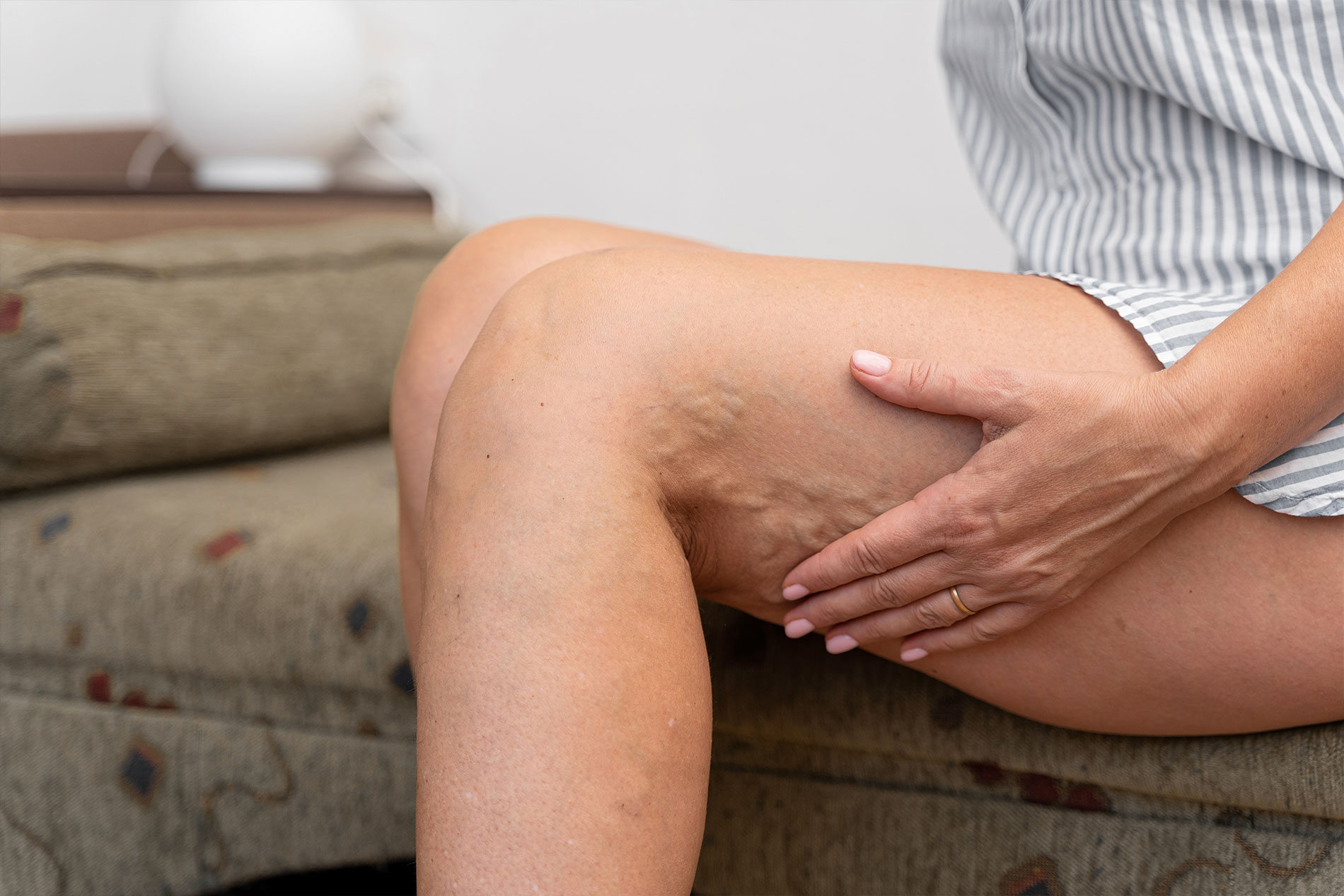
When you are diagnosed with Peripheral Arterial Disease (PAD), the medical terminology can feel overwhelming. Terms like “stenosis,” “occlusion,” and “revascularization” get thrown around, often leaving patients confused about their actual treatment options. At the heart of the matter is a simple problem: the arteries in your legs are blocked by plaque, and blood cannot get through. This lack of blood flow causes pain when walking (claudication), leg cramps, and in severe cases, non-healing wounds that can lead to amputation.
The solution seems straightforward—unclog the artery. However, the method used to achieve that goal is a subject of intense medical research and clinical expertise. Two of the most common minimally invasive procedures used today are angioplasty and atherectomy. Often, patients will hear their vascular specialist recommend one, or increasingly, a combination of both.
This raises a critical question for anyone facing a procedure: Is it better to just stretch the artery open (angioplasty), or is it superior to physically remove the plaque first (atherectomy)? The answer isn’t a simple yes or no; it depends on the nature of your blockage, the location of the disease, and the characteristics of the plaque itself. This comprehensive guide will break down the mechanics of these procedures, compare their effectiveness, and help you understand why a personalized approach from a vascular specialist is the key to long-term success.
The Basics: Understanding the Procedures
Before comparing them, it is essential to understand exactly what happens inside your body during these treatments. Both are “endovascular” procedures, meaning they are performed from the inside of the blood vessel. Dr. Fox performs these in an outpatient setting, inserting a thin tube called a catheter through a tiny puncture in the groin or wrist to reach the blockage. There are no large incisions, and recovery is typically rapid.
What is Angioplasty?
Angioplasty (specifically Percutaneous Transluminal Angioplasty, or PTA) is the grandfather of endovascular therapy. It has been the standard of care for decades.
- The Mechanism: A catheter tipped with a deflated balloon is guided to the narrow spot in the artery. Once positioned, the balloon is inflated to a high pressure.
- The Effect: The force of the balloon pushes the plaque outward against the artery wall. It essentially squashes the blockage to make the channel wider, restoring blood flow.
- The Limitation: Imagine trying to stretch a hard, calcified pipe. Sometimes the plaque is too hard to be pushed aside. Other times, the artery wall stretches but then snaps back (elastic recoil) once the balloon is deflated. Additionally, the stretching force can sometimes cause a small tear (dissection) in the artery wall, which requires a metal stent to fix.
What is Atherectomy?
Atherectomy takes a different approach. Instead of just pushing the plaque aside, it physically removes it. The name comes from “atheroma” (plaque) and “ectomy” (surgical removal).
- The Mechanism: A specialized catheter is used, equipped with a cutting, grinding, or sanding device at the tip.
- Directional Atherectomy: Uses a blade to shave plaque off the wall.
- Rotational Atherectomy: Uses a spinning burr to grind hard calcium into microscopic particles.
- Orbital Atherectomy: Uses an eccentrically mounted crown to sand away plaque.
- Laser Atherectomy: Uses high-energy light to vaporize the blockage.
- The Effect: By debulking the lesion (removing the mass), the channel is physically opened. The debris is either trapped in a filter basket downstream or aspirated (sucked out) through the catheter.
The Debate: Angioplasty Alone vs. Combination Therapy
For years, angioplasty alone was the go-to treatment. If the balloon didn’t work perfectly, a metal stent was placed to hold the artery open. While this saved many legs, it wasn’t a perfect solution. Stents in the legs—especially in areas that bend like the knee—can fracture or kink. Furthermore, the body sometimes reacts to the stent by growing scar tissue, leading to re-narrowing (restenosis).
This led to the rise of “vessel prep” strategies, where atherectomy is used before angioplasty. The theory is logical: if you remove the bulk of the plaque first, the balloon doesn’t have to work as hard, and you might not need a stent at all.
Why Combine Them?
When Dr. Fox uses atherectomy followed by angioplasty, the goal is to optimize the result.
- Debulking: Atherectomy removes the hard calcium and bulk.
- Compliance: This makes the artery more flexible (“compliant”).
- Gentle Expansion: Because the plaque is gone, the subsequent angioplasty balloon can be inflated at a lower pressure. This reduces trauma to the vessel wall, lowers the risk of tearing (dissection), and minimizes recoil.
- Better Drug Delivery: If a drug-coated balloon (DCB) is used, removing the plaque first allows the medication to penetrate deeper into the vessel wall, preventing future scar tissue.
Clinical Scenarios: When is Combination Therapy Better?
Not every blockage needs atherectomy. For soft, fresh plaque or simple narrowings, angioplasty alone (often with a drug-coated balloon) provides excellent results. However, there are specific scenarios where adding atherectomy clearly provides a superior outcome.
1. Heavily Calcified Arteries
This is the most significant differentiator. Calcium is like rock; a balloon cannot squash rock. If you try to expand a heavily calcified artery with a balloon alone, the balloon might rupture, or the artery might tear in an uncontrolled way.
- The Advantage: Atherectomy (specifically rotational or orbital) is designed to modify this calcium. It cracks the rock or sands it down, changing the compliance of the vessel so the balloon can effectively open it.
2. “No-Stent” Zones
Certain areas of the leg, specifically the popliteal artery (behind the knee) and the common femoral artery (in the groin), undergo tremendous movement. Every time you walk, sit, or bend your leg, these arteries twist and compress.
- The Problem: Placing a metal stent in these areas is risky because the metal can fracture or damage the artery over time.
- The Advantage: Atherectomy combined with angioplasty allows the surgeon to open the vessel without leaving permanent metal behind. This “leave nothing behind” strategy is crucial for younger patients or active individuals.
3. Complex or Long Blockages
Some patients have blocked leg arteries where the occlusion extends for several inches (long lesions).
- The Problem: Angioplasty alone on a long blockage has a high rate of failure and restenosis. The more surface area you stretch, the more trauma you cause.
- The Advantage: Debulking the long segment with atherectomy first reduces the plaque burden significantly, leading to better flow rates and durability than ballooning alone.
4. Total Occlusions
In some cases, the artery is 100% blocked.
- The Advantage: Crossing a total blockage and treating it often requires removing the material to create a channel, rather than just forcing a channel through the solid mass.
The Risks: Is More Always Better?
If atherectomy is so effective, why not use it on everyone? As with any medical procedure, there is a risk-benefit calculation. Adding atherectomy to the procedure adds cost, time, and specific risks.
- Distal Embolization: The process of grinding or shaving plaque creates debris. While protection devices (filters) are used to catch this debris, there is a small risk that microscopic particles could travel downstream and block the tiny vessels in the toes (trash foot). This risk is higher with atherectomy than with angioplasty alone.
- Vessel Perforation: Because atherectomy involves cutting or sanding, there is a slightly higher risk of accidentally damaging the artery wall compared to a balloon.
However, in the hands of an experienced operator like Dr. Fox, these risks are minimized through precise technique and advanced imaging guidance.
Measuring Success: What Does the Data Say?
Clinical studies comparing the two approaches have shown mixed but promising results for combination therapy in specific groups.
- The DEFINITIVE LE Study: This landmark trial showed that directional atherectomy was safe and effective for treating PAD, with high rates of keeping the artery open (patency) without the need for stents.
- Drug-Coated Balloons (DCB): Recent data suggests that the “synergy” of atherectomy plus DCB is powerful. The atherectomy preps the vessel, and the drug-coated balloon delivers paclitaxel (a medication) to stop cell overgrowth. This combination has shown superior results in calcified arteries compared to DCB alone.
Essentially, for simple “soft” plaque, angioplasty is often sufficient. But as the disease becomes more complex—more calcium, longer blockages, tougher locations—the addition of atherectomy becomes statistically vital for keeping the artery open long-term.
The Role of the Vascular Specialist: Personalized Care
This brings us to the most important point: PAD is not a generic disease. Your blockage is unique to your anatomy, your genetics, and your lifestyle. This is why you cannot rely on a one-size-fits-all approach.
A board-certified vascular specialist brings judgment to the table. Dr. Fox does not simply decide to use atherectomy because it is available; he uses it when the angiogram reveals specific features (like heavy calcium) that predict angioplasty alone will fail.
The Diagnostic Process
Before any wire crosses a blockage, extensive planning occurs.
- Non-Invasive Testing: In our vascular lab, we use ultrasound and ABI testing to understand the hemodynamic impact of the blockage.
- Angiography: During the procedure, the dye injection reveals the “morphology” of the plaque. Is it smooth? Irregular? Calcified? Eccentric (on one side)?
- Device Selection: Based on this real-time visual data, Dr. Fox selects the right tool. He might choose:
- Angioplasty Alone: For a short, focal narrowing.
- Atherectomy + Angioplasty: For a calcified artery behind the knee.
- Stenting: As a “bailout” if the other methods don’t achieve a perfect result.
Why “Amputation Prevention” is the Ultimate Goal
Whether we use a balloon, a laser, or a diamond-tipped burr, the goal remains the same: amputation prevention.
When arteries are blocked, the tissue downstream begins to die. This starts with pain and can end with gangrene. By restoring straight-line flow to the foot, we provide the oxygen and nutrients needed to heal wounds and keep the tissue alive.
Both angioplasty and atherectomy are tools of “limb salvage.” In the past, if angioplasty failed, a patient might have been told there was nothing else to do, or offered a major bypass surgery. Today, the ability to combine these technologies means we can treat patients with complex, severe disease in an outpatient setting, saving limbs that might otherwise have been lost.
Recovery and Long-Term Maintenance
Regardless of whether you have angioplasty alone or combined with atherectomy, the recovery is similar. Because these are minimally invasive PAD treatments, you go home the same day. There are no stitches, just a small bandage. You can typically walk immediately and return to normal activities within a day or two.
However, the durability of the result depends heavily on you.
- Smoking Cessation: Continuing to smoke is the fastest way to clog the artery again, regardless of how well the procedure went.
- Walking: A structured walking program helps maintain the flow we restored.
- Medical Management: Taking your antiplatelet and cholesterol medications is crucial.
Conclusion: Which is Better?
So, which works better: Atherectomy with Angioplasty or Angioplasty alone?
The answer is: The one that fits your anatomy.
- Angioplasty alone is excellent for simpler, softer, shorter blockages. It is less invasive, faster, and carries lower risk of embolization.
- Atherectomy + Angioplasty is superior for calcified, complex, or long blockages, and for areas where we want to avoid stents (like the knee). It prepares the vessel for a better, more durable expansion.
You do not need to make this decision yourself. You need a partner in your vascular health who has mastery of both techniques. At Fox Vein and Vascular, Dr. Fox utilizes the full spectrum of endovascular technology to ensure that every patient receives the specific treatment their arteries require. We don’t just treat the image on the screen; we treat the patient, ensuring the best possible chance for a pain-free, active life.
If you are experiencing leg pain, cramping, or have been told you have poor circulation, don’t wait until the options become limited. Early evaluation offers the widest range of minimally invasive solutions.
Schedule a consultation with Dr. Fox at Fox Vein and Vascular to discuss your personalized PAD treatment plan. Contact us today at (212) 362-3470 or visit foxvein.com to take the first step toward better vascular health.
Leading Manhattan Vascular & Vein Specialist
At Fox Vein Care, we provide state-of-the-art vascular and venous treatments, combining advanced diagnostic technology with minimally invasive procedures that prioritize comfort, safety, and outstanding results.
Contact Us