


Dr. Fox is an outstanding surgeon. Him and his staff explains everything clearly which makes for a smoother process. I've recommended him to family and several close friends, and they've all had the same excellent experience. If you want top-tier expertise with genuine compassion, Dr. Fox is the best. Highly recommend!
I had a very positive experience at Fox Vein Care. The doctor took time to explain everything clearly and made me feel comfortable throughout the process. The staff was also friendly and helpful. Overall, I felt well taken care of and am happy with my results. Highly recommended!
I had a great experience at this doctor's office. The doctor treated me with genuine care and respect, was very patient, and took the time to truly listen to my concerns. He explained everything clearly and gave thoughtful, helpful advice. I never felt rushed, and I left feeling confident and well taken care of. Highly recommend.
Dr. Fox is outstanding. He explains everything patiently and well. His staff is both efficient and kind. I wouldn't go anywhere else for vascular issues.
I have been a patient of Dr. Fox since he was at Roosevelt Hospital on 59th Street in Manhattan. He's the best, hands down! And his Staff are the best at what they do, along with excellent bedside manners. Crystal is the Greatest!

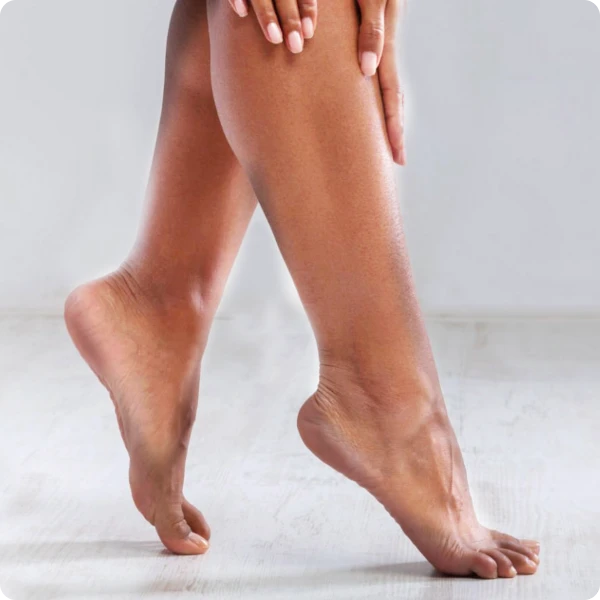
Before

After

Before

After

Before

After

Before
