If you’re dealing with uncomfortable or unsightly vein issues—from bulging veins and persistent vein pain to chronic leg swelling—your search for a “vein specialist near me” will likely lead to a recommendation for a vein ultrasound exam. This critical diagnostic step can feel mysterious or even a bit intimidating if you don’t know what to expect. However, this simple, non-invasive test is the most important tool a specialist has for understanding the root cause of your vein problems and designing an effective treatment plan.
A vein ultrasound, also known as a duplex ultrasound or venous doppler study, is the gold standard for evaluating the health of your veins. It provides a detailed, real-time map of your circulatory system, allowing your doctor to see what’s happening beneath the surface of your skin. This guide will demystify the entire process, explaining why the exam is necessary, how to prepare, what happens during the test, and what the results mean for your “vein care treatment in Manhattan“. Understanding this procedure is the first step toward getting the right diagnosis and finding lasting relief.
If you want an overview of advanced vein treatments in Manhattan and more information about the full range of services available beyond the ultrasound exam, check out our comprehensive vein treatment page.
You can also explore more about our specialized vein treatments and how we approach conditions like varicose veins and chronic venous insufficiency.
Why is a Vein Ultrasound Necessary?
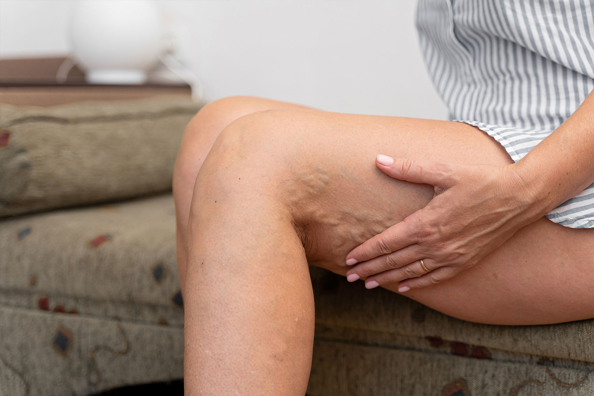
Many people assume that if they have visible varicose veins or spider veins, the problem is purely cosmetic and confined to the surface. In reality, these visible signs are often just the tip of the iceberg. They are typically symptoms of an underlying medical condition called chronic venous insufficiency (CVI).
Understanding Chronic Venous Insufficiency (CVI)
Your leg veins contain a series of one-way valves designed to help push blood upward toward the heart, against the pull of gravity. When these valves become weak or damaged, they fail to close properly. This allows blood to leak backward and pool in the lower legs, a condition known as venous reflux.
This pooling of blood increases the pressure inside the veins, causing them to stretch, twist, and bulge. This process leads to the full spectrum of “vein symptoms in legs”, including:
- Varicose Veins: Enlarged, rope-like veins.
- Leg Swelling and Veins: Edema in the ankles and calves, especially at the end of the day.
- Vein Pain: Aching, throbbing, cramping, or a feeling of heaviness.
- Skin Changes: Discoloration, itching, dryness, or even the formation of venous ulcers (open sores).
Because the underlying cause of these symptoms—venous reflux—is invisible to the naked eye, a specialist needs a way to see inside your veins. This is where the ultrasound exam becomes indispensable.
Learn more about how our diagnostic vascular laboratory supports accurate vein evaluations and tailored treatment plans.
The Purpose of the Exam
A “varicose vein doctor Manhattan” uses the ultrasound for several critical purposes:
- To Diagnose Venous Reflux: The primary goal is to identify which veins have faulty valves and to measure the severity of the backward blood flow. This confirms the diagnosis of CVI.
- To Create a “Vein Map”: The exam produces a detailed map of your unique venous anatomy. It shows the specialist the exact location, size, and path of the diseased veins, which is essential for planning a precise treatment like Endovenous Laser Therapy (EVLT).
- To Rule Out Other Conditions: The ultrasound is also used to check for more serious issues, most notably a deep vein thrombosis (DVT). A DVT is a blood clot in one of the deep veins of the leg. This is a potentially life-threatening condition that requires immediate and different medical treatment. Finding or ruling out a DVT is a crucial part of the initial evaluation.
- To Guide Treatment: During minimally invasive procedures, the specialist uses live ultrasound imaging to guide catheters and needles to their exact targets, ensuring the treatment is both safe and effective.
Without the detailed information provided by an ultrasound, any treatment would be mere guesswork. The exam ensures that the care you receive from a “Manhattan vein clinic“ is based on a precise, accurate diagnosis.
How to Prepare for Your Vein Ultrasound
One of the best aspects of a vein ultrasound is that it requires very little preparation on your part. It is a simple and straightforward procedure. However, following a few guidelines can help ensure the process goes smoothly and the results are as accurate as possible.
- Wear Comfortable, Loose-Fitting Clothing: You will likely be asked to change into a medical gown or shorts for the exam. Arriving in clothes that are easy to remove and put back on is helpful. Avoid tight-fitting pants or girdles.
- Stay Hydrated: Drinking a good amount of water in the 24 hours leading up to your exam can be beneficial. Well-hydrated veins are slightly larger and easier for the technologist to visualize and assess.
- Avoid Body Lotion: Do not apply any lotions, oils, or creams to your legs on the day of the exam. These products can interfere with the ultrasound gel and make it difficult for the transducer to get good contact with your skin.
- Bring Your Medical Information: Have a list of your current medications and be ready to discuss your symptoms and medical history with the technologist and doctor.
- No Fasting Required: Unlike some other medical tests, you do not need to fast before a vein ultrasound. You can eat and drink normally.
The staff at the clinic will provide you with any specific instructions you need. Don’t hesitate to ask questions if you are unsure about any aspect of the preparation. For more preparation tips, review our patient resources.
The Step-by-Step Vein Ultrasound Procedure
When you arrive at the “best vein doctor near me” for your exam, you will be guided through a process that is designed to be as comfortable and thorough as possible. The exam is typically performed by a Registered Vascular Technologist (RVT), a professional specifically trained in vascular ultrasound, and the results are then interpreted by the vein specialist. The entire exam usually takes between 30 to 90 minutes, depending on whether one or both legs are being evaluated.
Step 1: The Initial Conversation and Changing
The technologist will greet you, confirm your identity, and briefly discuss the symptoms you have been experiencing. They will explain the procedure to you and answer any initial questions you might have. You will then be given privacy to change into a gown or shorts, leaving your lower body accessible for the exam.
Step 2: Getting into Position
For a complete and accurate study, the technologist needs to evaluate your veins while you are in different positions. Most of the exam is performed while you are standing on a small, stable platform with a support rail to hold onto.
Why standing? Gravity. When you stand, the full effect of gravity on the blood in your leg veins can be observed. This position makes it much easier to detect venous reflux, as the backward flow of blood through faulty valves becomes more pronounced. A vein ultrasound performed only while lying down is incomplete and can miss the diagnosis. You may also be asked to lie down on an examination table for part of the test to evaluate the deep veins.
Learn more about our approach to diagnosis and evaluation.
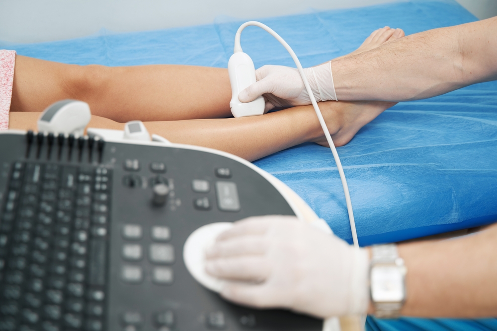
Step 3: The Ultrasound Gel and Transducer
The technologist will apply a clear, water-based gel to your skin over the areas to be examined. This gel might feel a bit cool, but it is harmless. Its purpose is to eliminate any air pockets between the handheld ultrasound probe (called a transducer) and your skin, ensuring the sound waves can travel effectively to create a clear image.
The technologist will then press the transducer firmly against your skin and move it along the path of your major veins, from the groin all the way down to the ankle.
Step 4: The Imaging and Doppler Assessment
As the technologist moves the transducer, images of your veins will appear on a monitor. The “duplex” part of the ultrasound comes from its two functions:
- Imaging (B-mode): The transducer sends out high-frequency sound waves that bounce off the tissues and veins in your leg. The returning echoes are used to create a black-and-white, two-dimensional image of the vein’s structure. The technologist can see the vein’s size, depth, and whether there are any visible blockages like a clot.
- Doppler: This is the part of the exam that assesses blood flow. The Doppler ultrasound measures the speed and direction of moving blood cells. This information is often represented by colors on the screen (color Doppler) and an audible “whooshing” sound (spectral Doppler). Blue typically indicates blood flowing away from the probe, while red indicates blood flowing toward it.
Step 5: The Augmentation Maneuvers
To test your vein valves, the technologist needs to see how they respond under pressure. They will perform a series of “augmentation” maneuvers. This is the most active part of the test. The most common maneuver is a calf squeeze.
The technologist will gently but firmly squeeze your calf muscle. This action forces blood to move up the leg. They will then quickly release the squeeze. In a healthy vein, the valves will snap shut, preventing any blood from flowing backward. The Doppler will show a brief rush of upward flow, followed by silence.
If you have venous reflux, the valves will not close properly. When the technologist releases the squeeze, the Doppler will detect blood flowing back down the leg. The technologist will measure the duration of this backward flow. Reflux lasting longer than 0.5 seconds is considered abnormal and confirms a diagnosis of CVI. This process will be repeated at several points along the main superficial veins, such as the great saphenous vein (GSV) and small saphenous vein (SSV).
To learn more about our doctors’ expertise in ultrasound-guided vein procedures, visit the about the specialist page.
Step 6: Completing the Exam
Once the technologist has gathered all the necessary images and measurements from your superficial and deep vein systems, the exam is complete. You will be given a towel to wipe off the gel, and you can get dressed. The procedure is entirely external, with no needles or radiation involved. There are no side effects, and you can immediately return to your normal daily activities.
Interpreting Your Ultrasound Results
After your exam, the vein specialist will meticulously review all the images and data collected by the technologist. They will analyze the findings to form a complete picture of your venous health. The report will detail:
- Deep Vein Patency: It will confirm whether your deep veins are “patent,” meaning they are open and free of any clots (DVT). This is a critical safety check.
- Superficial Vein Reflux: It will identify which specific superficial veins (like the GSV) have incompetent valves and are the source of the reflux.
- Vein Diameters: The report will note the diameters of the diseased veins. This information helps in planning treatments like EVLT.
- Tributary Varicosities: It will map out the branching, bulging veins that are connected to the main refluxing source veins.
The specialist will then sit down with you to discuss these findings in a way that is easy to understand. This is a crucial part of the service at a top chronic vein condition specialist. They will use the ultrasound results to explain exactly why you are having symptoms and recommend a personalized treatment plan designed to address the specific problems identified in your vein map.
Your Partner in Vein Health: Fox Vein and Vascular
Choosing where to have your vein ultrasound is as important as the test itself. The quality of the equipment and the expertise of the technologist and physician are paramount for an accurate diagnosis. At Fox Vein and Vascular, we pride ourselves on our state-of-the-art, in-house vascular laboratory. Our team, led by Dr. David Fox, a board-certified vascular surgeon, has decades of experience in diagnosing and treating the full spectrum of vein disease.
We understand that a medical test can be daunting, and our team is committed to making your vein ultrasound exam a comfortable, informative, and stress-free experience. We ensure that you understand every step of the process and take the time to thoroughly review the results with you, empowering you to make confident decisions about your “vein treatment near me”.
A vein ultrasound exam is a painless, safe, and powerful diagnostic tool. It is the essential first step on your journey toward alleviating vein pain, reducing leg swelling, and eliminating varicose veins. By providing a clear window into your vascular system, the ultrasound allows your specialist to move beyond treating symptoms and instead focus on resolving the underlying cause of your vein disease, leading to effective, long-lasting relief.
For more information on all the conditions we treat and to learn about personalized treatment options, visit our Conditions We Treat page or contact us to schedule a consultation.
Leading Manhattan Vascular & Vein Specialist
At Fox Vein Care, we provide state-of-the-art vascular and venous treatments, combining advanced diagnostic technology with minimally invasive procedures that prioritize comfort, safety, and outstanding results.
Contact Us